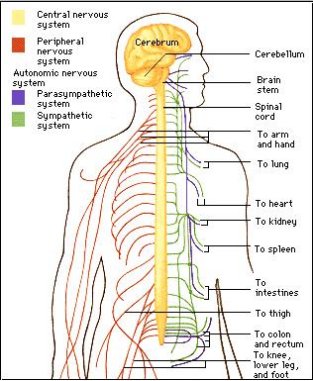
Organs of the nervous system
|
|
The Brain
The brain is vital to our existence. It is one of the most important organs in the human body, and controls everything we do, both voluntary and involuntary, and gives us things like emotions, memories and personality.
The brain is a very complex organ, a sort of body system in it's own right. It has taken scientists centuries to understand just some of the brain's many processes, and many of these still remain a secret. What we know for sure, is that it's the organ that makes us, giving people the ability to create art, learn a language, see the world, and have moral judgment and rational thought.
All this comes from a gelatinous mass of fats and proteins that takes up about 3 pounds of our body weight and uses one fifth of our oxygen and energy. While not sounding impressive, it is one of the body's biggest organs, containing some 100 billion nerve cells that not only put together thoughts and physical actions but also do things like regulate our breathing and keep our hearts beating.
The cells found in the brain are called neurons, which make up the organ's "gray matter." The neurons transmit and gather signals that are sent around via a network of millions of nerve fibers called dendrites and axons. These create the brain's "white matter."
*For more info on this, check out the Cells and Tissues of the Nervous System section of the website.
- The Cerebrum-The cerebrum is the largest part of the brain, taking up about eighty percent of it's weight. The deeply-wrinkled outer surface of the cerebrum is called the cerebral cortex, which is gray matter. Beneath it is the white matter. It's the cerebrum that makes the human brain so astounding. Other animals, like elephants, dolphins, and whales have much larger brains than we do, but the human brain contains a cerebrum that is the most developed of any animal. The cerebrum - which literally means "brain" in Latin-- is the most evolutionarily recent and largest part of the brain as a whole. Here, things like perception, imagination, thought, judgment, and decisions are made. The cerebral cortex is composed of six thin layers of neurons, which sit on top of thick white matter pathways. The cortex is heavily condensed, and if you were to lay it out flat, it would take up about 2 1/2 square feet. Within that, it includes about 10 billion neurons, and about 50 trillion synapses.
The folds in the cortex have ridges, called gyri, and valleys, which are called sulci. Normally, a sulcus is quite long and pronounced, and are boundaries between the four subdivisions of the cerebrum, called lobes.
The first is called the frontal lobe. This lobe is responsible for voluntary movements and planning, and scientists believe that it plays the most significant role in personality and intelligence. The second largest part of the brain is the cerebellum, which sits at the back of the cerebrum. It coordinates our muscle movements and controls balance. Having of both grey and white matter, the cerebellum transmits information to both the spinal cord and other parts of the brain. Thirdly, the diencephalon, which is in the brain's core. It is a complex of intricate structures that creates a mass roughly the size of an apricot, the two major sections being the thalamus and hypothalamus. The thalamus acts as a relay station for nerve impulses from around the body that are then sent to the correct part of the cerebrum to be processed. The hypothalamus controls hormone releases from the pituitary gland. These hormones control growth and instinctual behavior like eating. - The cerebrum has two halves, or hemispheres. It is further subdivided into four regions, or lobes, in each hemisphere. The frontal lobes, located at the forehead, are responsible for speech, thought, learning, emotion, and movement. At the back portion of the frontal lobe is an area called the motor cortex. In studies using brain surgery patients, electrical signals were sent to the motor cortex, causing body parts to move. This has allowed for researchers to actually map out the motor cortex based on what parts it controls very precisely. The lowest portions of the motor cortex control the muscles of the mouth and face, while areas near the top of the head control the legs and feet. Behind them are the parietal lobes, which process information sent by sensory organs like touch, temperature, and pain. It includes an area called the somatosensory cortex. A similar test to the one conducted on the motor cortex also found that patients described sensations of being touched at various parts of their bodies. Just like the motor cortex, the somatosensory cortex can be mapped with the mouth and face closest to the temples and the legs and feet at the top of the head.
At the lower back of the brain are the occipital lobes, which controls vision, and lastly, there are the temporal lobes, near the temples, which are involved with hearing and memory. - The hemispheres-When a brain is veiwed from the top, it becomes immediately obvious that it is split in two from front to back. There are two hemispheres in our brains, almost as if we have two brains in our heads instead of just one. These two halves are linked together with white matter called the corpus callosum.
The two halves of the brain specialize in different areas. The left hemisphere generally controls the right side of the body, and the right hemisphere controls the left. Also, the left hemisphere that usually interprets language, and seems to be responsible for similar systems, like math and logic. The right hemisphere has more to do with things like spatial orientation, face recognition, and body image. It also governs our ability to appreciate art and music.
Some of the most interesting work done showing the differences of the two hemispheres was done by Roger Sperry. He worked with people who had had operations to control severe epilepsy. In some cases, severe epilepsy can be almost completely eliminated by cutting out parts of the corpus callosum. In a sense, these people really did have two different brains.
Sperry found that if he put something in the right hand of one of these people after they had their operation, they could communicate what it was, But if he put it in their left hand, they would know what is was, but were not able to say what it was. Why did this happen? When something is held in the right hand, it goes to the left hemisphere and, since that's the side with language, the person could say what it was. The feeling of the thing in the left hand, though, went to the right hemisphere, and since parts of the corpus callosum were removed, and the right hemisphere is only in charge of remembering names, the two hemispheres could not communicate.
In the occipital lobe, the eyes are hooked up to the hemispheres in a somewhat complicated way: The right hand side of the retinas (which sees things to the left of a focal point) goes to the right hemisphere, and the left hand side of each retina (which see things to the right) goes to the left hemisphere. What this means is that, if you have someone stare at a point and briefly show them something on the left, the right hemisphere will receive the information. If you show them something on the right, it is the left hemisphere that receives the information.
Sperry would flash things on a projection screen and ask the patients to either say what they saw or pick what they saw with one hand or the other from a box full of things. So, if he showed a ball on the left side of the screen and a pencil on the right, the person would say "pencil" (using the speech centre on the left side) but pick a ball from the box with their left hand using the right side.

|
|
- The brain stem, located below the cerebrum, controls reflexes and basic life functions, like heart rate, breathing, and blood pressure, and also regulates when you feel sleepy or awake. The brain is extremely sensitive and delicate, and needs maximum protection, provided by the skull and three tough membranes called meninges. The spaces between the meninges, the blood-brain barrier, are filled with fluid that cushions the brain and keeps it from being damaged by contact with the inside of the skull.
The spinal Cord
The spinal cord is the main pathway for information between the nervous system and the brain. The human spinal cord is protected by the bony spinal column, made up of bones called vertebrae. The spinal column is somewhat flexible, some of the vertebrae in the lower parts of the spinal column become fused.
The spinal cord is made up of 31 segments: 8 cervical, 12 thoracic, 5 lumbar, 5 sacral and 1 coccygeal. A pair of spinal nerves are found at each segment of the spinal cord, with different amounts of gray and white matter. The spinal cord is about 43 to 45 centimeters, and is slightly shorter than the spinal column.
Nerves that extend from the spinal cord from the lumbar and sacral segments run in the vertebral canal for a distance before they leave the vertebral column. These nerves in the vertebral canal are called the cauda equinae, or horse tails.
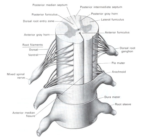
There are many parts to the spinal cord. Some of them are:
1. Posterior Median and Intermediate Steptum
2. Posterior/Anterior gray horn
3. Lateral/Anterior Funiculus
4. Dorsal Root Ganglion
5. Dura and Pia Mater
6. Arachnoid Nerve Branches
7. Nerve Root Sleeve
8. Anterior Median Fissure
9. Spinal Nerves
10. Dorsal and ventral root filaments
The spinal cord is made up of 31 segments: 8 cervical, 12 thoracic, 5 lumbar, 5 sacral and 1 coccygeal. A pair of spinal nerves are found at each segment of the spinal cord, with different amounts of gray and white matter. The spinal cord is about 43 to 45 centimeters, and is slightly shorter than the spinal column.
Nerves that extend from the spinal cord from the lumbar and sacral segments run in the vertebral canal for a distance before they leave the vertebral column. These nerves in the vertebral canal are called the cauda equinae, or horse tails.
There are many parts to the spinal cord. Some of them are:
1. Posterior Median and Intermediate Steptum
2. Posterior/Anterior gray horn
3. Lateral/Anterior Funiculus
4. Dorsal Root Ganglion
5. Dura and Pia Mater
6. Arachnoid Nerve Branches
7. Nerve Root Sleeve
8. Anterior Median Fissure
9. Spinal Nerves
10. Dorsal and ventral root filaments
